Microvisioneer mvSlide
Manual Whole Slide Scanner
Upgrade your microscope to a manual slide scanner.
Start creating virtual slides now for any digital pathology application.
Create unlimited panoramas with unmatched image quality, flexibility and scan speed.
mvSlide - The affordable real-time image stitching solution from the market leader - trusted by experts in 80+ countries worldwide.
Used in 100+ peer-reviewed publications from leading institutions.
mvSlide software


mvSlide
professional performance
panoramic image stitching
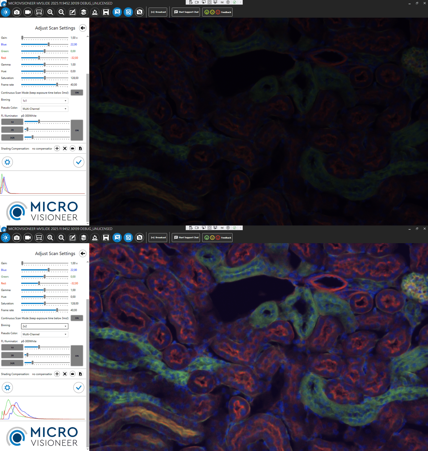
New mvSlide 2026 Fluorescence Edition available now -
featuring several upgrades including a highly requested pixel binning for higher sensitivity for low-intensity samples!


See How to Scan with mvSlide
Manual Scanning with mvSlide
Zoom into Scans Created with mvSlide
Scan Right Away on Your Desk -
Benefits of Microvisioneer mvSlide
Affordable
Upgrading of microscope with the mvSlide software is inexpensive. No maintenance costs occur. The manual scanner can be an alternative to an automated scanner or an add-on or back-up.
Time Saving
Immediately available solution.
Scanning of smaller areas is often faster compared to automated scanners.
Manual scanning compensates for outage of automated scanners.
Flexible
Maintains flexibility and features of the microscope.
Slides can still be assessed via the eyepiece.
Versatile
The manual scanning system is suitable for exceptional slides, such as thick or uneven slides.
Possibilities of Whole Slide Imaging with Microvisioneer mvSlide
Image Analysis
Use the hiqh-quality whole slide images to perform representative and reproducible analyses. Automate image analysis.
Documentation
Save your digital slides, annotations and comments.
Preserve the original staining.
Create publication-ready high-quality images.
Education
Scan your own teaching slides and move away from expensive glass slides.
All students see exactly the same virtual slide, remotely and simultaneously.
Collaboration
Share virtual microscope slides with your colleagues worldwide.
Work remotely and save time and costs.
Applications
Microvisioneer's manual scanning approach combines digital progress and innovation with the versatility and flexibility of traditional microscopes. With the resulting low cost manual microscope scanner, almost any type of slide can be digitized, no matter how exceptional.
Scan interesting slides right away on your desk, scan particularly thick slides, unusually large slides or uneven slides, which are often a problem for automated scanners.
Typical brightfield histology tissue slide scanning or cytology imaging, digitization of frozen sections, oil immersion, phase contrast, polarized light and fluorescence imaging is all possible. Set up your own histology or pathology scanner, tailored to your needs.
A broad range of cameras is supported, among others various Olympus / Evident DP cameras, e.g. DP27, DP28, DP74, DP75, or Carl Zeiss Axiocam cameras, e.g. AxioCam 305.
Explore a few selected applications of Microvisioneer manual scanning in more detail in the use case section on this website.
For research use only.

human intestine, H&E stain

gynecologic sample, papanicolaou stain

blood smear, Wright's stain

rock thin section

skin of pig embryo

BPAE cells stained with MitoTracker Red CMXRos and Alexa Fluor 488
Some of Our Customers


What Our Customers Say
“IT GIVES A CLEAR OVERVIEW. IT IS VERY FAST. IT HAS A GREAT RESOLUTION. IT IS EASY TO FOCUS WHEREEVER YOU NEED IT."
“A WORKING SCANNER THAT PRODUCES DIAGNOSTIC QUALITY IMAGES"
“I CAN USE MY EXISTING EQUIPMENT, NO NEED FOR A MAJOR CAPITAL INVESTMENT. MANY OF THE FEATURES ARE UNIQUE AND USEFUL."
“UNPRECEDENTED SMOOTHNESS IN THE IMAGE STITCHING, WHICH IS IMPRESSIVE."
“PRACTICAL AND EASY USABILITY FOR OIL-BASED SCANNING."


