
autoWSI
Motorized
Whole Slide Imaging
for Specialized Microscopy
Applications

Software-based WSI
Built around configurable motorized stages and suitable microscopes.
Motorized control
X-, Y and Z-axis workflow.
Preview scan and autofocus.
Flexible modalities
2X - 100X magnification.
Brightfield incl. oil immersion, polarized light - fluorescence coming soon.
See autoWSI scan performance
Watch autoWSI automatically digitize a large orangutan brain section, from preview image acquisition and focus map creation to motorized image acquisition and stitching.
What autoWSI offers
1
Macro and Label Image &
Preview Scan
autoWSI can be configured to acquire a slide image. In high-magnification workflows, overview scans help define scan areas.
2
Region Selection, Autofocus & Focus Mapping
Define focus points within the selected scan regions. autoWSI automatically determines the optimal focus and generates a focus map for automated z-axis adjustment during scanning.
3
Automated Scanning & Stitching
Scanning of selected regions or full slides and creation of whole slide images.
A configurable microscope-based automated system
Application examples for specialized motorized WSI

Geology, Geoarchaeology & Petrographic Materials
Thin sections and mineral-based samples often require microscope-specific contrast settings such as polarized light. autoWSI combines this optical flexibility with motorized acquisition, enabling high-resolution digitization of larger sample areas.
Typical examples:
Thin sections - Rocks - Sediments - Ceramic or mortar samples
Published research using autoWSI
Reconstructing human–animal interactions via multiproxy analysis of dog paleofeces from the medieval Central Balkans
Archaeological and Anthropological Sciences, 2026
Late Antique—Early Byzantine urban transformation in Mid-Northern Anatolia: A multiproxy approach from Pompeiopolis
PLoS One, 2026

High Magnification Oil Immersion Scanning
autoWSI can be configured for high-magnification workflows where manual scanning becomes inefficient, especially when larger regions or repeated scans are required.
Typical examples:
Blood smears - Bone marrow smears - Cytology - Detailed ROIs

Large Tissue Sections & Scan Areas
autoWSI automates image acquisition and stitching, making digitization of large sections more efficient. For non-standard slide formats, autoWSI can be configured with suitable stage travel, sample holder and scanning parameters.
Typical examples:
Brain sections - Large tissue sections - Non-standard slide formats
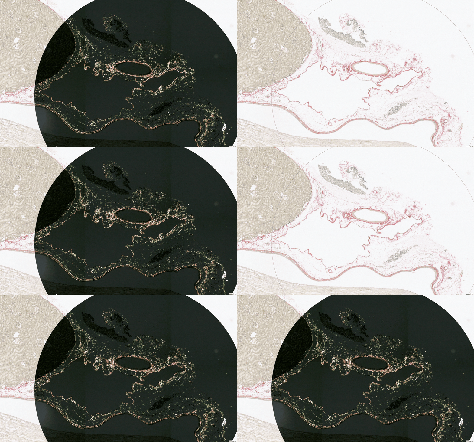
Polarized Light Imaging in Histological Research
Histological applications such as Picrosirius Red or Congo Red staining benefit from polarized light imaging, which is not supported by many conventional brightfield scanners. By utilizing the polarization capabilities of a suitable microscope, autoWSI enables automated scanning of these samples.
Typical examples:
Picrosirius Red stained collagen fibers - Congo Red stained amyloid deposits
For research use only.
Why choose a modular scanning solution based on autoWSI?
autoWSI is designed for research laboratories that need motorized whole slide imaging while keeping the optical and application-specific flexibility of a microscope-based setup

Upgrade an Existing Microscope
A suitable microscope can be upgraded with a motorized stage, camera and autoWSI software to enable automated whole slide scanning.

Go Beyond Standard Scanning
Make use of microscope-specific optics and contrast methods to scan samples that conventional slide scanners may not accomodate - including polarized light, oil-immersion and non-standard slide formats.

A cost-efficient Alternative for Specialized WSI
By reusing suitable microscope hardware, autoWSI can offer a more affordabe path to specialized motorized scanning than dedicated high-end scanning systems.
SIGN UP, DISCUSS YOUR SETUP AND RECEIVE A QUOTE
autoWSI systems are configured according to the microscope, motorized stage, camera and intended application. Please provide a few details about your setup and scanning requirements so that we can assess the most suitable configuration and prepare initial advice or a quotation.
Microvisioneer GmbH


